Followers

Sunday, April 21, 2019

Alzheimer’s disease

Alzheimer’s disease

Dementia is a syndrome characterized by progressive loss of previously acquired cognitive skills including memory, language, insight, and judgment.
Alzheimer's disease accounts for the majority (50% to 75%) of all cases of dementia.
Approximately 10% of all persons over the age of 70 have significant memory loss, and in more than half the cause is AD

EPIDEMIOLOGY & DEMOGRAPHICS

•INCIDENCE: Risk doubles every 5 yr after the age of 65; above the age of 85th incidence is about 8%.
•PREVALENCE: Currently an estimated 4 million Americans have AD; 7% between the ages of 65 and 74, 53% between the ages of 75 and 84, and 40% over the age of 85.
•PREDOMINANT SEX: Female


The Brain and Alzheimer’s Disease

Two major structural changes:

1.Neurofibrillary tangles-Bundles of twisted threads that are the product of collapsed neural structures (contain abnormal forms of tau protein
2.Amyloid plaques-Dense deposits of deteriorated amyloid protein, surrounded by clumps of dead nerve and glial cells.

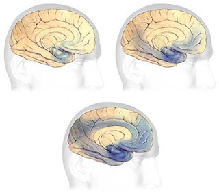
earliest and most severe degeneration is usually found in the medial temporal lobe
: (entorhinal/perirhinal cortex hippocampus),
   lateral temporal cortex, nucleus basalis of Meynert
These images represent a cross-section of the brain as seen from the front. The cross-section on the left represents a normal brain and the one on the right represents a brain with Alzheimer's disease.In Alzheimer's disease, there is an overall shrinkage of brain tissue. The grooves or furrows in the brain, called sulci (plural of sulcus), are noticeably widened and there is shrinkage of the gyri (plural of gyrus), the well-developed folds of the brain's outer layer. In addition, the ventricles, or chambers within the brain that contain cerebrospinal fluid, are noticeably enlarged. In the early stages of Alzheimer's disease, short-term memory begins to fade (see box labeled ‘memory') when the cells in the hippocampus, which is part of the limbic system, degenerate. The ability to perform routine tasks also declines. As Alzheimer's disease spreads through the cerebral cortex (the outer layer of the brain), judgment declines, emotional outbursts may occur and language is impaired. As the disease progresses, more nerve cells die, leading to changes in behavior, such as wandering and agitation. In the final stages of the disease, people may lose the ability to recognize faces and communicate; they normally cannot control bodily functions and require constant care. On average, the disease lasts for 8 to 10 years, but individuals with Alzheimer’s can live for up to 20 years.

Severe Alzheimer’s Disease


Several competing hypotheses:

Cholinergic hypothesis
-Caused by reduced synthesis of acetylcholine
-Destruction of these neurons causes disruptions in distant neuronal networks (perception, memory, judgment)
Amyloid hypothesis
-Abnormal breakdown; buildup of amyloid beta deposits
-Damaged amyloid proteins build to toxic levels, causing call damage and death
Tau hypothesis
-Caused by tau protein abnormalities
-Formation of neurofibrillary tangles

PHYSICAL FINDINGS & CLINICAL PRESENTATION
Family member, not the patient, often notes insidious memory impairment.    
Patients have difficulties learning and retaining new information, handling complex tasks (e.g., balancing the checkbook), and have impairments in reasoning, judgment, spatial ability, and orientation (e.g., difficulty driving, getting lost away from home). 
Behavioral changes, such as mood changes and apathy, may accompany memory impairment.
In later stages patients may develop agitation and psychosis.

DIAGNOSIS

Diagnosis is dependent on clinical history, a thorough physical and neurologic examination, and use of reliable and valid diagnostic criteria (i.e., DSM-IV or NINDCS-ADRDA) such as the following:
  Loss of memory and one or more additional cognitive abilities (aphasia, apraxia, agnosia)
Insidious onset and gradual progression of symptoms
Impairment in social or occupational functioning
 Cognitive loss documented by neuropsychologic tests  
 No physical signs, neuroimaging, or laboratory evidence of other diseases that can cause dementia (i.e., metabolic abnormalities, medication or toxin effects, infection, stroke, Parkinson's disease, subdural hematoma, or tumors)
There is no definitive imaging or laboratory test for the diagnosis of dementia.
NINCDS–ADRDA :National Institute of Neurological Disorders and Stroke—Alzheimer’s Disease and Related Disorders Association
DSM-IV :Diagnostic and Statistical Manual of Mental Disorders

DIFFERENTIAL DIAGNOSIS

Cancer (brain tumor, meningeal neoplasia)
Infection (AIDS, neurosyphilis, progressive multifocal leucoencephalopathy)
Metabolic (alcohol, hypothyroidism, B12 deficiency)
Organ failure (dialysis dementia, Wilson's disease)
Vascular disorder (chronic subdural hematoma)
Depression

WORKUP

HISTORY & GENERAL PHYSICAL EXAMINATION:   
Medication use should always be reviewed for drugs that may cause mental status changes.  
Patients should be screened for depression, because it can sometimes mimic dementia but also often occurs as a coexisting condition and should be treated.
On examination, look for signs of metabolic disturbance, presence of psychiatric features, or focal neurologic deficits.
MENTAL STATUS TESTING:
Brief mental status testing.
Most commonly used is the Folstein Mini-Mental Status Examination (MMSE). A MMSE score <24 (scores range from 0 to 30, with lower scores reflecting poorer performance) suggests dementia;
MMSE is not sensitive enough to detect mild dementia,
dementia in patients with high baseline IQ. Scores may be spuriously low in patients with limited education, poor motor function, poor language skills, or impaired vision.
Attention is usually preserved until the late stages of AD, so consider alternate diagnoses in patients who do poorly on tests of attention.

LABORATORY TESTS

CBC
Serum electrolytes. BUN/creatinine Glucose , Liver and thyroid function testsSerum vitamin B12 and methylmalonic acidSyphilis serology, if high clinical suspicionLumbar puncture if history or signs of cancer, infectious process, or when the clinical presentation is unusual (i.e., rapid progression of symptoms)
EEG if there is history of seizures, episodic confusion, rapid clinical decline, or suspicion of Creutzfeldt-Jakob disease
Measurement of apolipoprotein E genotyping, CSF tau and amyloid,
Functional imaging including positron emission tomography (PET) or scanning proton emission computed tomography (SPECT) are not routinely indicated
IMAGING STUDIES
CT scan or MRI to rule out hydrocephalus and mass lesions, including subdural hematoma.

TREATMENT

NONPHARMACOLOGIC THERAPY
Patient safety, including risks associated with impaired driving, wandering behavior, leaving stoves unattended, and accidents, must be addressed with the patient and family early and appropriate measures implemented.
Wandering, hoarding or hiding objects, repetitive questioning, withdrawal, and social inappropriateness often respond to behavioral therapies.
ACUTE GENERAL Rx
None
CHRONIC Rx
1.    Symptomatic treatment of memory disturbance:a.    Cholinesterase inhibitors (ChEI): for mild to moderate AD (MMSE 10-26). Common side effects include nausea, diarrhea, and anorexia.b.    NMDA (N-methyl-D-aspartate )receptor antagonist: Memantine). for moderate to severe AD. Common side effects include constipation, dizziness, or headache. Memantine is contraindicated in patients with renal insufficiency or history of seizures.
2.    Symptomatic treatment of neuropsychiatric and behavioral disturbances: Depression, agitation, delusions, or hallucinations.

Wednesday, April 17, 2019

Aplastic Anemia

Aplastic Anemia

Deficiency or injury to the stem cells
Hypocellular bone marrow
Results in pancytopenia of the peripheral blood
All cell lines affected

        Severity

Moderate aplastic anemia
Marrow cellularity <30%
Absence of severe pancytopenia
Depression of at least two of three blood elements below normal.
Severe
oBone marrow cellularity <25% or marrow showing <50% normal with two of three peripheral blood count criteria:  
oANC <500
Plt <20k
Retic count <40k
Very Severe
All of above plus ANC less than 200.
Classification
Inherited
Fanconi’s anemia, dyskeratosis congenita, Shwachman-Diamond Syndrome, Reticular dysgenesis, Amegakaryocytic thrombocytopenia, familial aplastic anemia, preleukemia (monosomy 7) and nonhematologic disease (Down, Dubowitz, Seckel)
Acquired
Irradiation
drugs and chemicals: cytotoxic agents, benzene, idiosyncratic reaction, chloramphenicol, NSAIDS, antiepileptics, Gold
viruses: EBV, Hepatitis virus (non-A,non-B, non-C, non-G), Parvovirus (transient aplastic crisis or pure red cell aplasia), HIV
Immune diseases: eosinophilic fasciitis, hyperimmunoglobulinemia, thymoma and thymic carcinoma, GvHD in immunodeficiency
PNH
Pregnancy
Idiopathic 

Differential Diagnosis

Pancytopenia with hypocellular bone marrow
Acquired aplastic anemia  - Inherited aplastic anemia
Hypoplastic MDS  - Hypoplastic AML
Pancytopenia with cellular bone marrow
Primary bone marrow diseases  -MDS
PNH  - Myelofibrosis
Myelophthisis  - Bone marrow lymphoma
Hairy cell leukemia  - SLE, Sjogren’s disease
Hypersplenism  - Vitamin B12 and folate deficiency
Overwhelming infection  - Alcoholism
Brucellosis  - Ehrlichiosis
Sarcoidosis  - tuberculosis
Hypocellular bone marrow with or without cytopenia
Q fever  - Legionaires disease
Mycobacteria  - Tuberculosis
Hypothyroidism  - Anorexia nervosa

Etiology

   —RadiationChemicalsDrugsInfectionsImmunological  diseasesPregnancyParoxysmal nocturnal hemoglobinuriaConstitutional disorders

Drugs causing aplastic anemia

Cytotoxic drugs like alkylating agents, antimitotics
Benzene
Low probability : chloramphenicol,insecticides,antiprotozoals,  NSAIDS, anticonvulsants,heavy metals,

Pathogenesis

Genetic predisposition found in HLA-DR2. 
-This correlates to response to immunosuppressants.
Drug Injury
Immune mediated injury
Immune-mediated T-cell destruction of marrow
Removal of lymphocytes from aplastic bone marrow improved colony number in tissue culture and addition of lymphocytes to normal marrow inhibited hematopoiesis in vitro.

Presenting Symptoms

       —Related to anemiaFatigue, Shortness of breathRelated to neutrapeniaInfectionsRelated to thrombocytopeniaBleeding (mucous membranes)


Diagnosis

Blood macrocytic picture with thrombocytopenia and low granulocytes
Reticulocytes are reduced or few
No immature cell
Bone marrow:-
Ancillary studies: - chromosomal breakage study,flow cytometry, serological studies to exclude viral infections, MRI  spine

Bone marrow in aplastic Anemia

Marrow is profoundly hypocellular with decrease in all elements.
Residual hematopoietic cells are morphologically normal.
Malignant infiltrates and fibrosis is absent.
Hematopoiesis is non-megaloblastic.


Treatment 

SupportiveBlood product transfusion & antibiotics1/3 refractory to plateletsBleeding deaths uncommon

Definitive Treatment

Bone marrow transplantation
Immunosuppression no difference in long-term survival

Definitive Treatment

Immunosuppression Older patientsNo sibling donorsAntithymocyte globulin (ATG) +/- cyclosporineResponse rate = 70%
Long term survival = 65-90% 
Marrow Transplantation
Genotypically identical sibling donor marrow
Cures aplastic anemia
Death can occur as a result of complications of the procedure
Long-term survival with younger pts 65%

Tuesday, April 16, 2019

THALASSEMIA SYNDROMES

THALASSEMIA SYNDROMES

The thalassemia syndromes are inherited disorders of α- or ß-globin biosynthesis.

 The reduced supply of globin diminishes production of hemoglobin tetramers
synthesis of the unaffected globins proceeds at a normal rate. Unbalanced accumulation of α and ß subunits causing hypochromia and microcytosis.

CLINICAL MANIFESTATlONS OF  THALASSEMIA SYNDROMES


Mutations causing thalassemia can affect any step in the pathway of globin gene expression:Transcriptionprocessing of the mRNA precursor translationPost-translational metabolism of the ß-globin
 The most common forms arise from mutations that derange splicing of the mRNA precursor or prematurely terminate translation of the mRNA.
Hypochromia and microcytosis reduced amounts of hemoglobin tetramers
in heterozygotes (-thalassemia trait), this is the only abnormality seen. Anemia is minimal.
 In more severe homozygous states, unbalanced globin accumulation
 causes accumulation of highly insoluble unpaired chains.
They form toxic inclusion bodies that kill developing erythroblasts in the marrow. 
The surviving RBCs bear a burden of inclusion bodies that are detected in the spleen
 shortening the RBC life span and producing severe hemolytic anemia.
The resulting profound anemia stimulates erythropoietin release and compensatory erythroid hyperplasia
Erythroid hyperplasia can become exuberant and produce masses of extramedullary erythropoietic tissue in the liver and spleen.
Massive bone marrow expansion deranges growth and development.
Children develop characteristic "chipmunk" facies due to maxillary marrow hyperplasia and frontal bossing.
Thinning and pathologic fracture of long bones and vertebrae may occur due to cortical invasion by erythroid elements and profound growth retardation.


Hemolytic anemia causes hepatosplenomegaly, leg ulcers, gallstones, and high-output congestive heart failure.
Chronic transfusions with RBCs improve oxygen delivery, suppress the excessive ineffective erythropoiesis, and prolong life
iron overload, usually prove fatal by age 30 years.
When both  alleles on one chromosome are deleted, the locus is called -thal-1; when only a single  allele on one chromosome is deleted, the locus is called -thal-2.
90–95% of the hemoglobin is hemoglobin Barts (tetramers of chains).-Thalassemia-2 trait is an asymptomatic, silent carrier state.-Thalassemia-1 trait resembles -thalassemia minor. 

HbH disease

HbA production is only 25–30% normal.
Fetuses accumulate some unpaired chains (Hb Barts; -chain tetramers).
 In adults, unpaired chains accumulate and are soluble enough to form 4 tetramers called HbH.
 HbH forms few inclusions in erythroblasts and precipitates in circulating RBC.
 Patients with HbH disease have thalassemia intermedia characterized by moderately severe hemolytic anemia but milder ineffective erythropoiesis.
 Survival into midadult life without transfusions is common.

HYDROPS FETALIS

The homozygous state for the -thalassemia-1 cis deletion (hydrops fetalis) causes total absence of -globin synthesis.
No physiologically useful hemoglobin is produced beyond the embryonic stage.
 Excess globin forms tetramers called Hb Barts (4), which has a very high oxygen affinity.
 It delivers almost no O2 to fetal tissues, causing tissue asphyxia, edema (hydrops fetalis), congestive heart failure, and death in utero.

Diagnosis and Management of Thalassemias

The diagnosis of -thalassemia major is readily made during childhood on the basis of severe anemia accompanied by the characteristic signs of massive ineffective erythropoiesis:
Hepatosplenomegaly
profound microcytosis
 a characteristic blood smear 
 and elevated levels of HbF, HbA2, or both.
Many patients require chronic hypertransfusion therapy designed to
maintain a hematocrit of at least 27–30% so that erythropoiesis is suppressed
Splenectomy is required if the annual transfusion requirement (volume of RBCs per kilogram of body weight per year) increases by >50%.
 Folic acid supplements may be useful.
 Vaccination with Pneumovax in anticipation of eventual splenectomy is advised
 close monitoring for infection, leg ulcers, and biliary tract disease.
 Many patients develop endocrine deficiencies as a result of iron overload
 Early endocrine evaluation is required for glucose intolerance, thyroid dysfunction, and delayed onset of puberty or secondary sexual characteristics.

thalassemia intermedia

Have similar stigmata but can survive without chronic hypertransfusion.
A number of factors can aggravate the anemia, including infection, onset of puberty, and development of splenomegaly and hypersplenism.
 Some patients may eventually benefit from splenectomy.

Thalassemia minor (i.e., thalassemia trait)

usually presents as profound microcytosis and hypochromia with target cells
only minimal or mild anemia.
The mean corpuscular volume is rarely >75 fL
hematocrit is rarely <30–33%.
 Hemoglobin analysis classically reveals an elevated HbA2 (3.5–7.5%), but some forms are associated with normal HbA2 and/or elevated HbF.
 Genetic counseling and patient education are essential.
Patients with -thalassemia trait should be warned that their blood picture resembles iron deficiency and can be misdiagnosed.
 They should eschew empirical use of iron, yet iron deficiency can develop during pregnancy or from chronic bleeding .
Persons with -thalassemia trait may exhibit mild hypochromia and microcytosis usually without anemia. HbA2 and HbF levels are normal.
 Affected individuals usually require only genetic counseling.
 HbH disease resembles -thalassemia intermedia, with the added complication that the HbH molecule behaves like moderately unstable hemoglobin.
 Patients with HbH disease should undergo splenectomy if excessive anemia or a transfusion requirement develops.
 Oxidative drugs should be avoided.
 Iron overload leading to death can occur in more severely affected patients.

Transfusional Hemosiderosis

Chronic blood transfusion can lead to bloodborne infection, alloimmunization, febrile reactions, and lethal iron overload .
 A unit of packed RBCs contains 250–300 mg iron (1 mg/mL).
The iron assimilated by a single transfusion of two units of packed RBCs is thus equal to a 1- to 2-year intake of iron.
 Iron accumulates in chronically transfused patients because no mechanisms exist for increasing iron excretion
 Vitamin C should not be supplemented because it generates free radicals in iron excess states.
Patients who receive >100 units of packed RBCs usually develop hemosiderosis.
The ferritin level rises, followed by early endocrine dysfunction (glucose intolerance and delayed puberty), cirrhosis, and cardiomyopathy.
Liver biopsy shows both parenchymal and reticuloendothelial iron.
 The superconducting quantum-interference device (SQUID) is accurate at measuring hepatic iron but not widely available. Cardiac toxicity is often insidious.
Early development of pericarditis is followed by dysrhythmia and pump failure.
 The onset of heart failure is ominous

CHELATING AGENTS

 long-term transfusion support prompts one to institute therapy with iron-chelating agents.
Desferoxamine (Desferal) is for parenteral use.
Its iron-binding kinetics require chronic slow infusion via a metering pump.
 The constant presence of the drug improves the efficiency of chelation and protects tissues from occasional releases of the most toxic fraction of iron—low-molecular-weight iron.
Desferoxamine is relatively nontoxic.
Occasional cataracts, deafness, and local skin reactions, including urticaria, occur. Skin reactions can usually be managed with antihistamines.
Negative iron balance can be achieved, even in the face of a high transfusion requirement.
To enjoy a significant survival advantage, chelation must begin before 5–8 years of age in -thalassemia major.
Deferasirox is an oral iron-chelating agent.
Single daily doses of 20 to 30 mg/kg deferasirox produced reductions in liver iron concentration comparable to desferoxamine in long-term transfused adult and pediatric patients.
Deferasirox produces some elevations in liver enzymes and slight but persistent increases in serum creatinine, without apparent clinical consequence.
 Other toxicities are similar to those of desferoxamine.
 Its toxicity profile is acceptable, although long-term effects are still being evaluated

Experimental Therapies

Bone Marrow Transplantation, Gene Therapy, and Manipulation of HBF
Bone marrow transplantation provides stem cells able to express normal hemoglobin; it has been used in a large number of patients with  thalassemia and a smaller number of patients with sickle cell anemia.
 Early in the course of disease, before end-organ damage occurs, transplantation is curative in 80–90% of patients.
 In highly experienced centers, the treatment-related mortality is <10%.
Gene therapy of thalassemia and sickle cell disease has proved to be an elusive goal.